Breast Thermography is an assessment tool we offer at Thermography Clinic Ireland. It is a safe, non-ionizing, non-contact study of breast skin temperature that is useful as a breast health risk assessment. It has particular utility in monitoring the effects of breast hormone therapy. It is also useful as in the detection of physiologic changes such as hormonal imbalance, lymphatic edema, ductal congestion, chest wall pain syndromes, and angiogenesis. Internationally peer reviewed Guidelines for Breast Thermography have been developed by the American Academy of Thermology in 2012 and restated in 2013. These guidelines support breast thermal imaging as a breast risk assessment, not unlike high blood pressure screening for vascular disease.
Thermography measures, images and maps microcirculatory shunting associated with breast circulatory changes in the skin. Sex hormones, in particular estrogen and progesterone, can affect breast physiology and circulation. Serial studies are helpful in monitoring the effects of hormone replacement therapy and the treatment of fibrocystic disease. There are several musculoskeletal applications that impact breast lymphatics, health and associated pain as well.
The role of Thermography in cancer detection is frequently misunderstood and deserves special attention.
Angoiogensis has been called a breast risk health assessment as cancer cells need increased blood flow (angiogenesis) in order to “take” over surrounding breast tissue. They also have an increased metabolic rate, which translates into an increase in temperature compared to surrounding normal tissue. By studying breast tissue with infrared imaging early changes in blood flow can be detected and progressive changes can be assessed over time.
In 1997 Gamagami, Silverstein & Waisman published that
- Angiogenesis was the first sign appearing on mammography before the appearance of image of breast cancer, predicting in 91 % of the cases which breast might develop breast carcinoma. This is an important finding in the detection of the early stages of breast cancer development.
- Infrared imaging goes hand in hand with mammography. Hypervascularity and hyperthermia could be shown in 86% of non-palpable breast cancer. In 15% it helped to detect the cancer upon an unsuspicious image on mammography.
- Infrared imaging was found to be the only test showing the efficiency of chemotherapy in inflammatory breast carcinoma.
There was also a 95% negative predictive value, and a 24% positive predictive value. It has been argued that this means if an Infrared Thermogram is negative there is a 95% chance that there is no cancer and that if it is positive than there is a 24% chance that cancer may later be discovered.
Infrared Thermography is a valuable adjunct to X-ray mammography and ultrasound, especially in women with dense breast tissue. Other indications include small breasts, post implant breasts, post mastectomy breasts, those looking for additional information, and to follow the effects of treatment.
Thermal imaging is an examination of physiology that is complimentary to anatomical imaging techniques. Although proven to be highly accurate, thermal imaging is an adjunctive procedure; and as such, it is not intended to replace anatomic studies such as mammography, ultrasound, MRI, CT, X-ray, or others.
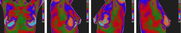
Example Breast Thermography Protocol Images: