News

Breast Cancer Prevention Beyond the Mammogram

This article underscores the importance of not relying on just one modality to protect us from cancer.  Although mammography has been a powerful tool in the fight to detect breast cancer early, it is not the only tool we have and combining a variety of diagnostic and preventative tools is truly the best way to avoid invasive breast cancer.

  1. Mammography. This test is currently the standard of care for screening for breast cancer.  But women must remember that this is an x-ray of the breast.  The breast is not an air-filled cavity or a bone so the pictures that are generated are limited.  The mammogram is limited by dense breasts, meaning that a small cancerous tumor can be hidden in-between breast tissue.  Also, keep in mind that this procedure exposes breast tissue to trauma and radiation, both of which can be cancer causing to this delicate tissue.
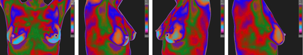
  2. Thermography-a picture generated by a heat sensing camera.  A “hotspot” in the breast can be indicative of angiogenesis, which is the formation of new blood vessels to feed the developing tumor.  This is a great way to make healthy lifestyle changes before cancer actually forms or to remove the cancer before it has a chance to spread at all.  Breast thermography is not limited by density of breast tissue, making it an excellent test for younger women (20-50 years old).  Thermography is underutilized due to its lack of regulation and general acceptance by the medical community. Thermography is intended to be an adjunctive test, not to stand alone from mammography. Thermograms are sometimes done with improper cameras or with improper follow-up.  That is why we offer thermography in our office.  Our camera is top of the line,
  3. Self-Breast Exam.  Most of the women that I know who have had breast cancer found it themselves.  We should never underestimate the power of knowing our own bodies.  Women should be encouraged from a young age to know their breasts normal lumps and bumps.   When they are older, they will be confident that they will notice any changes in their breasts.
  4. Prevent breast cancer by breastfeeding and healthy lifestyle choices like exercise and low sugar diets.  Studies have shown that the breast is changed by the breastmilk flowing through it.  There is actually a component of breastmilk that has an anti-tumor function.  The greatest benefit is achieved from breastfeeding when a woman was breastfed as a child and then goes on to breastfeed her offspring.  Exercise has been shown to decrease risk for almost all diseases including breast cancer.  Sugar causes inflammation and acidity in our bodies; cancer cells love this type of environment and this chemistry change can make a woman’s immune system less able to recognize and eliminate cancer cells (source).

Remember that breast cancer is not limited to women with a family history, all women must be aware of the risk of breast cancer and work toward prevention. Call to day to schedule your Breast Thermography Appointment. Remember Breast Thermography is a non-invasive, radiation-free assessment tool

Breast Thermography

Providing a risk assessment for a health condition offers a patient so much more than just limited disease detection. There are many conditions that currently have biomarkers that will warn far in advance of significant pathology. It is far better to warn of impending heart disease and diabetes than to suddenly find you have it and that significant damage has already been done. These risk markers give you time to make changes to either prevent the condition or at least control it. Thermography brings this same risk assessment to the breast health of every woman [4,5]. Not only can breast thermography warn that a cancer may be present, but also provide a marker of future risk along with a role in prevention [3-5, 10-12].

The use of thermography is based on the principle that metabolic activity and vascular circulation in both pre-cancerous tissue and the area surrounding a developing breast cancer is almost always higher than in normal breast tissue. In an ever-increasing need for nutrients, cancerous tumors increase circulation to their cells by holding open existing blood vessels, opening dormant vessels, and creating new ones (neoangiogenesis). This process frequently results in an increase in regional surface temperatures of the breast. Thermography uses ultra-sensitive medical infrared cameras and sophisticated computers to detect, analyze, and produce high-resolution images of these temperature variations. Because of thermography’s detection ability, these temperature variations may be among the earliest signs of breast cancer and/or a pre-cancerous state of the breast [3,6-9].

Current methods used to detect suspicious signs of breast cancer depend primarily on the combination of both physical examination and mammography. While this approach has become the mainstay of early breast cancer detection, more is needed. Since the absolute prevention of breast cancer has not become a reality as of yet, efforts must be directed at detecting breast cancer at its earliest stage. As such, the addition of thermography to the frontline of early breast cancer detection brings a great deal of good news for women.

WHAT MAKES INFRARED IMAGING SO UNIQUE 

While mammography, ultrasound, MRI, and other types of structural imaging rely primarily on finding the physical tumor, thermography is based on detecting the heat produced by increased blood vessel circulation and metabolic changes associated with a tumor’s genesis and growth. By detecting minute variations in normal blood vessel activity, infrared imaging may find thermal signs suggesting a pre-cancerous state of the breast or the presence an early tumor that is not yet large enough to be detected by physical examination, mammography, or other types of structural imaging [3,6-9].

Certain types of cancers will not be detected (approximately 20%) by mammography for various reasons [9], but some of these cancers will be discovered by thermography [3,6-9].

Difficulties in reading mammograms can occur in women who are on hormone replacement, nursing or have fibrocystic, large, dense, or augmented breasts (6,8). These types of breast differences do not cause difficulties in reading infrared images.

THERMOGRAPHY AS A RISK MARKER FOR BREAST CANCER

Studies show that an abnormal infrared image is the single most important marker of high risk for developing breast cancer, 10 times more significant than a family history of the disease [5]. Consequently, in patients with a persistent abnormal thermogram, the examination results become a marker of higher future cancer risk [4-5]. Depending upon certain factors, re-examinations are performed at appropriate intervals to monitor the breasts. This gives a woman time to take a pro-active approach by working with her doctor to improve her breast health. By maintaining close monitoring of her breast health with infrared imaging, self breast exams, clinical examinations, structural imaging, and other tests, a woman has a much better chance of detecting cancer at its earliest stage and preventing invasive tumor growth.

Angiogenesis, or new blood vessel formation, is necessary to sustain the growth of a tumor. Thermography may be the first signal that such a possibility is developing [3].

Just as unique as a fingerprint, each patient has a particular infrared map of their breasts. Any modification of this infrared map on serial imaging (images taken over months to years) may constitute an early sign of an abnormality. However, if a pathology is suspected, this information is used to recommend further examinations and tests. 

THERMOGRAPHY’S ROLE IN BREAST CANCER PREVENTION

In another and possibly critical role, infrared imaging of the breast may hold a significant potential in breast cancer prevention. Due to the ability of infrared imaging’s detection of changes in the dermal circulation, any external pharmacological intervention or release of biochemicals by the body that have the propensity to alter blood flow may be detected. This is especially true of chemicals that are target specific for the tissues of the breast. Being a primary target tissue for the hormone estrogen, the hormone’s effect in the breast is anabolic to the ductal cells. As such, the outcome is one of increased cellular metabolism. This increase in cellular activity necessitates the need for nutrients above and beyond the norm. In order to facilitate this need, an increase in blood supply must occur. This translates to an infrared image demonstrating a uniform increase in vascular patterning (see accompanying images).

The importance of this observation lies in one of the primary risk factors for breast cancer—lifetime exposure to estrogen. If infrared imaging has the ability to warn of increased thermovascular activity due to increased levels of estrogen in the breast (estrogen dominance), action can be taken to lower this activity and ultimately the patient’s risk for future breast cancer. Treatments can be monitored for positive effects by incorporating infrared imaging as a method of observing these effects. Studies have shown this effect and the positive outcome of pharmacological intervention. Many patients with this condition also demonstrate signs and symptoms that include breast pain, tenderness, cysts, and benign lumps. In many patients, a reversal or reduction in these signs and symptoms are also noted when treatment is initiated [10–12]. Infrared imaging’s ability to detect increased thermovascular activity secondary to levels of estrogen in the breast, and to monitor the effects of treatment targeted at the breast, may play a significant role in breast cancer prevention.

TECHNOLOGY, INTERPRETATION AND COMPARATIVE IMAGING

As is the case with mammography and all other imaging modalities, access to sophisticated technology and the expertise to interpret the findings are of prime importance. To help distinguish a normal process from an abnormal one requires proper training, clinical experience, strict adherance to guidelines and protocols, and meticulous image acquisition. While not all tumors are visible on a mammogram, not all tumors are associated with a high level of blood vessel activity; thus, escaping infrared detection. Less aggressive lesions can be associated with less evident images. Therefore, in these select cases, thermography may be an indicator suggesting a much better overall prognosis.

When digitally produced, and interpreted by qualified doctors, abnormalities or changes in infrared images provide invaluable information. This is particularly true in patients with dense breasts, non-specific physical or mammographic findings, or women with a previous history of breast surgery or radiation. The use of serial infrared imaging can draw additional attention to areas that require further evaluation or closer scrutiny during initial or subsequent exams. This further evaluation may include additional structural imaging. Used as a complimentary imaging technique, recent data suggests that thermography may also help monitor the effects of some of the newer proposed anti-angiogenesis therapies (currently recognized as a promising treatment strategy) [6,8].

Since we have not been able to prevent breast cancer as of yet, there is a consensus among experts that more lives will be saved with earlier detection. Since both physical and mammographic examination cannot detect all cancers, particularly smaller tumors in younger patients and those with dense breast tissue, there is currently much interest in finding new ways to improve our abilities in early detection. While some techniques have emerged, most are designed to be used in selected cases where physical and mammographic examinations have already picked up an abnormality. Consequently, we are left with some patients who have adhered to current screening guidelines and are still left with undetected breast cancer. Therefore, experts have concluded that no one procedure or method of imaging is solely adequate for breast cancer screening [1,2,6].

EARLY DETECTION MEANS LIFE

Breast cancer is the most common cancer in women, and the risk increases with age [1]. Risk is also higher in women whose close relatives have had the disease. Women without children, and those who have had their first child after age 30, also seem to be at higher risk. However, every woman is at risk of developing breast cancer. Current research indicates that 1 in every 8 women in the US will get breast cancer in their lifetime [1].

Studies show an increase in survival rate when breast thermography and structural imaging are used together [3].

Thermography’s ability to detect thermal signs that may suggest a pre-cancerous state of the breast, or signs of cancer at a very early stage, lies in its unique capability of monitoring the temperature variations produced by the earliest changes in tissue physiology (function) [3,6-9]. However, no single imaging technology can offer 100% detection of all early stage cancers.  As such, if we are to offer women a method of true early detection, thermography, mammography, ultrasound, and MRI cannot be used alone. These are all adjunctive imaging technologies. Consequently, thermography’s role is in addition (an adjunct) to structural imaging and physical examination, not in lieu of. Thermography does not replace any other imaging technology, nor can any other imaging technology replace thermography. The tests complement each other. Since it has been determined that 1 in 8 women will get breast cancer, we must use every means possible to detect cancers when there is the greatest chance for survival. What we are promoting is a multimodal (multiple tests) approach to screening. The combined use of breast self-exams, physician exams, thermography, and structural imaging together provide the earliest detection system available to date [3,7-9]. If treated in the earliest stages, cure rates greater than 95% are possible [3,6]. With thermography’s expanded role as a risk assessment technology, infrared imaging brings a great deal of good news to women. 

WHO SHOULD HAVE THIS TEST?

Every woman should include breast thermography as an addition to her regular breast health care. We must do everything we can to provide earliest detection in an effort to prevent women from having to deal with this terrible disease.

Breast cancers are particularly aggressive in younger women. Statistics indicate that approximately 15% of all breast cancers occur in women under the age of 45 [13]. However, there are no clear guidelines for the use of imaging procedures during these years. With the addition of breast thermography, women in this age group have a tool that they can add to their regular breast health check-ups.

REFERENCES

  • 1. American Cancer Society – Breast Cancer Guidelines and Statistics, 2009-2010
  • 2. I. Nyirjesy, M.D. et al; Clinical Evaluation, Mammography and Thermography in the Diagnosis of Breast Carcinoma. Thermology, 1986; 1: 170-173.
  • 3. M. Gautherie, Ph.D.; Thermobiological Assessment of Benign and Malignant Breast Diseases. Am. J. Obstet. Gynecol., 1983; V 147, No. 8: 861-869.
  • 4. C. Gros, M.D., M. Gautherie, Ph.D.; Breast Thermography and Cancer Risk Prediction. Cancer, 1980; V 45, No. 1: 51-56.
  • 5. P. Haehnel, M.D., M. Gautherie, Ph.D. et al; Long-Term Assessment of Breast Cancer Risk by Thermal Imaging. In: Biomedical Thermology, 1980; 279-301.
  • 6. P. Gamigami, M.D.; Atlas of Mammography: New Early Signs in Breast Cancer. Blackwell Science, 1996.
  • 7. J. Keyserlingk, M.D.; Time to Reassess the Value of Infrared Breast Imaging? Oncology News Int., 1997; V 6, No. 9.
  • 8. P. Ahlgren, M.D., E. Yu, M.D., J. Keyserlingk, M.D.; Is it Time to Reassess the Value of Infrared Breast Imaging? Primary Care & Cancer (NCI), 1998; V 18, No. 2.
  • 9. N. Belliveau, M.D., J. Keyserlingk, M.D. et al ; Infrared Imaging of the Breast: Initial Reappraisal Using High-Resolution Digital Technology in 100 Successive Cases of Stage I and II Breast Cancer. Breast Journal, 1998; V 4, No. 4
  • 10. Verzini, L., Romani, L., Talia, B., (Radiology department university of Modena (Italy)). Thermographic variations in the breast during the menstrual cycle. Acta Thermographica, 143–149, 1980s.
  • 11. Huber, C., Pons, J., Pateau, A., (Gynecology and department of radiology Cretei hospital (Paris) France). Breast fibrocystic disease and thermography. Acta Thermographica, 48–50, 1980s.
  • 12. Borten, M., Ransil, B. et al. (Department of obstetrics and gynecology, Beth Israel Hospital, Harvard Medical School). Regional differences in breast surface temperature by liquid crystal thermography. Thermology, 1, 216–220, 1986.
  • 13. American Cancer Society – Breast Cancer Guidelines and Statistics

Consistency is Key but Sometimes is Better than Not At All

Tips to Help Improve Breast Health

  • Consider your toxic chemical load from your environment.  Limit the use of plastic and what chemicals are in the products you use to clean your home and what you apply to your skin. Reduce exposure to xenoestrogens. A large number of synthetic chemical compounds that we all encounter have estrogen-like activity.  Among them: common pesticides, industrial pollutants and hormone residues in meat, poultry and dairy products.
  • Do not keep your cell phone in your bra!  Several studies show the damage it causes. Plus, what is it doing to your heart and thyroid being so close to them.  Avoid carrying it on your body when possible.
  • Consider turning off wifi on phone, e-reader, or tablet when reading a book or playing games.  Think about where it’s resting while you are in bed.

Limit bra wearing.  Switch to underwire free if possible.  Decrease time wearing any bra.  The softer less constricting the better. Or none at all!

  • Skin brushing to move lymph.  Up towards heart and star pattern from nipple out over breasts.
  • Good nutrition-avoid sugar and limit consumption of high calorie low nutritional foods.
  • Eat more of a variety of vegetables and fruits, especially cruciferous.  They help reduce damaging estrogen and boost beneficial estrogen. They contain cancer fighting nutrients, 8-10 servings a day. Broccoli sprouts are a great addition to sandwiches, salads or just a snack
  • Spinach, kale, romaine lettuce, Swiss chard, and other dark green vegetables. They also boast plenty of carotenes, along with fiber and foliate. Two large studies suggest a relationship between increased folate (not folic acid that’s a synthetic form) intake and decreased breast cancer risk.
  • Garlic, onions, scallions, leeks, and other vegetables from the allium family.  Studies in animals show that allium vegetable components can slow the progression of several cancers, including breast cancer. 
  • Beans — lentils, peas, kidney beans, navy beans, and so on. Beans are rich in fiber, and also in an antioxidant that appears to slow tumor growth.
  • Adequate fiber daily will be beneficial, at least 28 grams/day. Lots of benefits, but it helps flush bile that grabs excess estrogen.
  • Limit consumption of highly processed meats.  Grass fed hormone free when possible.  Cooking to well done is more carcinogenic than medium. 
  • Choose fats wisely.  Extra virgin and cold pressed quality olive oil, organic saturated animal fat, increase omega-3 fats, found in cold-water fish (especially wild salmon and sardines), freshly ground flaxseed and walnuts have also been associated with inhibiting the growth of breast tumors.

Drink green tea. Regular consumption of green tea has been linked to a lower incidence of many kinds of cancer.

  • If consumed at all, limit alcohol consumption to one drink per day or less.
  • Switch to sea salt.  Limit eating salty foods and avoid foods processed with iodized salt.
  • Be as lean as possible without becoming underweight. (Fat stores estrogen.)
  • Add quality essential oils to your routine like Frankincense, Lavender, Clary Sage, and Sandalwood (I trust Young Living brand and can be a source for you if you need one).
  • Balance hormones and avoid long term birth control pills especially under age 18 and avoid synthetic hormone replacement therapy. 
  • Breastfeeding your children decreases the risk of breast cancer.  The longer the better.
  • Stop smoking.
  • Reduce your stress and support your emotions any way you can.  Yoga, exercise, meditate, laugh with friends, deep breathing, relaxing baths with Epsom salt, use essential oils, etc.
  • Get good sleep– melatonin may have antioxidant properties that help prevent damage to cells. In addition, melatonin lowers estrogen production from the ovaries.  Poor sleep alters hormones that influence cancer cells as well as the body not being able to do its repair work during sleep.
  • Exercise-be physically active for 20 minutes every day.  1.3 hours/week (Moves lymph, helps with estrogen, insulin handling and stress reduction.)
  • Give your liver some attention.  It has to help with hormones and detoxifying.  If it is congested, it taxes the body. 

4 mocktail recipes for sober October!

Published on  by Laura Southwood

You’ve likely heard of dry January, but have you heard of sober October? Consuming alcohol regularly may increase your risk of developing breast cancer. Here are some of my favorite mocktail recipes:

We all know that taking a break from alcohol can have positive effects on our health. With alcohol-free months like dry January and sober October increasing in popularity, there are more alcohol-free drink options available nowadays than ever before! Whether you’re looking to cut down on alcohol or go sober this October, read on for some mocktail inspiration.

Elderflower, cucumber & lime mocktail

With this zesty mocktail, you won’t feel like you’re missing out on anything! If you’re feeling fancy, why not substitute the elderflower for alcohol-free prosecco?

Ingredients (serves 2-4) –

  • 1/2 cucumber, diced
  • A small handful of fresh mint + extra for garnishing
  • 500ml sparkling elderflower
  • Juice of 2 limes
  • Ice (for serving)
  • Stevia (to sweeten – optional)

Method –

Place the cucumber, mint, lime juice and a big splash of (tap) water into a blender and blitz until smooth. You’ll need enough water for the ingredients to blend well, but not too much to make it too runny. Sieve the mixture into a jug, pressing those last bits through with the back of a spoon. Add the sparkling elderflower to taste and stir. Pour into your glass of choice, adding an ice cube or two and mint to garnish.

Blackberry & apple ‘lemonade

If – like us – you’ve been foraging for blackberries recently, this recipe is a great way to use up your surplus and add to your 5 (ahem, 10-) fruit and veg a day!

Ingredients (serves 4) 

  • 500g blackberries (fresh or frozen)
  • 1 apple, cored & diced
  • Juice of 2 lemons
  • 300ml tonic water or lemonade
  • Ice (for serving)

Method –

Place the blackberries, diced apple (no need to peel), lemon juice and a big splash of (tap) water into a blender and blitz until smooth. You’ll need enough water for the ingredients to blend well, but not too much to make it too runny. Strain through a sieve into a serving jug, add in the tonic water and stir to combine. Serve over ice. Top tip – save the pulp and add it to your yoghurt or cereal as a fruity topper the next morning!

Side note: any berries will work! Read more about the health benefits of fresh vs frozen foods here.

(Alcohol-free!) Bloody Mary

Did you know that there are numerous health benefits to a Bloody Mary? This alcohol-free version is a meal unto itself and is rich in nutrients such as fibre, vitamins and antioxidants.2

Ingredients (serves 2- 4) 

  • 200ml tomato juice
  • 5 splashes of Tobasco sauce (adjust to suit)
  • 2 tsp Worcestershire Sauce
  • Juice of 1/2 a lime
  • 1 stick of celery (to garnish)
  • Pinch of black pepper
  • Half a lime, cut into wedges + salt (for a salt rim – optional)

Method –

Cut the lime into two and chop one half into wedges. If you’re feeling fancy, moisten the rim of your serving glass(es) with 1 of the lime wedges and dip it in salt to create a salt rim. Combine the ingredients into a jug, stir and pour into your serving glasses. Garnish with celery, a wedge of lime and tuck in!

Long-Island Ice Tea

Research suggests that tea may be beneficial for your gut health by improving the composition of your gut microbiota!3 Not to mention apple cider vinegar may also help improve blood sugar regulation, weight loss, lower cholesterol and more!4

Ingredients (serves 4) 

  • 3 tea bags ( your tea of choice!)
  • 3 TBSP apple cider vinegar
  • 500ml ginger ale/beer
  • Juice of 2 lemons
  • Juice of 1 large orange (or 2 small!)
  • Mint sprigs, orange & lemon slices to garnish (optional)

Method –

Boil 3 cups of water and steep the 3 tea bags for 5-10 minutes. Discard the tea bags and leave the tea to cool. Once chilled, add the ACV, lemon juice and orange juice. Top with ginger ale or ginger beer and give it a stir. Serve with ice, orange/lemon slices and a few sprigs of mint.

Want to know more about the benefits of fermented food? Click here!

Click here to find out why we suggest sweetening with stevia Instead of sugar and for more gut-healthy drinks, check out a week’s worth of super supper smoothies for gut health

References

Before a Lump Develops

Alexander Mostovoy, H.D., D.H.M.S., B.C.C.T.

The statistics today are alarming: one in eight women will develop breast cancer. Each woman must choose between lumpectomy, mastectomy, radiation or chemotherapy in deciding how best to respond to her situation. With no cure for breast cancer in sight, everyone agrees that early detection is crucial.

Presently, breast self-examination and regular mammograms are the prescribed tools for early detection. Self-examination is an easy, no-cost way to monitor breast health and detect changes in the early stages. Mammograms provide a look inside the breast tissue and can often detect and diagnose anatomical abnormalities – such as lumps – already existing in the breast.

But lumps and other breast abnormalities generally don’t appear overnight. Long before they are discovered via film or fingers, their cellular contributors are hard at work. Cancer cells require a supply of blood in order to flourish and survive. Being aggressive, they create their own pathways to obtain this nourishment. Self-examination and mammograms cannot detect this invisible, early-cellular process, which usually occurs in breast tissue from five to ten years before even the slightest growth of a lump.

The science of breast thermography has opened the door to the earliest screening for abnormalities in breast tissue that Western medicine has ever known. Thermography uses infrared imaging to generate highly detailed digital pictures. The military began using infrared imaging in the 1950′s. Valued for its ability to detect and pinpoint areas of heat and movement, this technology provided a way for the military to “see’ what and where the eye or other surveillance techniques couldn’t -it is also known as “night vision”.

While the FDA has approved breast thermography since 1982, the greatest developments in this field have emerged in just the last few years with the advent of digital photography and high-resolution digital imaging. What is extraordinary about these intricate images is that they are able to display patterns of heat and vascularity within the tissue itself. Patterns of heat and vascularity can reveal the earliest indications of disease while they are still in the formative stages. Thermography can also detect the presence of heat from an infectious process or from a pre-existing anatomical abnormality (i.e. lump) in the breast. Thermography is also the best way to detect soft tissue trauma or damage. It is a non-invasive, radiation-free, safe and painless process.

The January 2003 issue of the American Journal of Radiology presented the results of a four-year clinical trial that tested thermographic imaging efficacy in distinguishing between benign and malignant lesions on patients who were scheduled for biopsies. Researchers concluded that, “infrared imaging is an economic and safe modality that provides physiological data about a lesion. The physiological view provided by infrared imaging complements the anatomical view provided by mammography… Thus, this … could be a valuable addition to the physician’s armamentarium of diagnostic tools.”

Breast thermography is not a stand-alone tool in the screening and diagnosis of breast cancer. It is adjunctive. We cannot ignore the tremendous role of thermography as an early-risk indicator or as a monitor of treatment. When a thermogram is positive, a closer look at the patient’s diet, exposure to environmental pollution, toxins and lifestyle is in order. Clinical blood work, in addition to ultrasound and mammography, is essential. When mammography and blood work is negative or equivocal, thermographic monitoring on a quarterly to semi-annual basis should be performed in those patients with suspicious thermograms.

Changes in tumor angiogenesis can be evaluated and other procedures can be ordered to aid in the earliest possible diagnosis. Thermography is non-ionizing and safe – there is no reason to simply “wait and see” any longer. It is at this stage that a major decision must be made. We can no longer be passive but must become proactive with our health. The “wait and see’ attitude due to a negative or equivocal mammogram is no longer acceptable. With a more universal and non-political approach, thermal imaging can become a valuable early-detection tool that is indispensable to a patient’s healthcare.

We must use every means possible to detect cancer where there is the greatest chance for survival. Proper use of breast self-exams, physician exams, thermography, and mammography together provide the earliest detection system available to date. If treated in the earliest stages, cure rates greater than 95% are possible.

Dr. Alexander Mostovoy is recognized as a leading authority on the application of clinical thermography. Since 1999, he has pioneered the use of Infrared Medical Thermography in his clinic in Toronto, Canada with a special interest in breast thermography and women’s health. I am priviledged to work with Dr Alex Mostovy & have his expertise & imput on my clients Breast Thermography reports.

Busting Cholesterol Myths with Carol Petersen RPh, CNP

Cholesterol-myths-with-carol-petersen-rph-Busting+Cholesterol+Myths+with+Carol+Petersen /thermographyclinic.com/blog

In our latest interview with Carol Petersen, RPh, CNP a compounding Pharmacist of many years, we busted the myth that cholesterol contributes to increased risk for cardiovascular disease:

·       Find out the importance of cholesterol to your hormone production and balance.

·       Understand the risks of suppressing cholesterol levels with statin drugs.

·       Learn about the most reliable test score when it comes to predicting the risk of heart disease.

·       Discover which key vitamins play a role in proper hormone function.

You don’t want to miss this interview where you will take away new knowledge and easy steps to implement toward bettering your health.

 You can watch it HERE: http://www.thermographyclinic.com/videos

error: Content is protected !!