News

Adrenal Fatigue and Cancer: Is There a Connection?

By Dr. Veronique Desaulniers

The worldwide low thyroid function epidemic is getting more and more press these days. But adrenal fatigue and its connection to major disease like cancer? Not so much. Research says there is definitely a link.

What are the Adrenals?
The adrenals are glands that rest just above the kidneys. They are the main production center for stress hormones, namely cortisol and adrenaline. What a lot of people don’t know, however, is that the adrenals also produce the reproductive hormone progesterone. According to many studies including a 2007 Italian investigation published in the journal Breast Cancer Research, progesterone is not only responsible for reproductive health. It is also a powerful cancer-protector.

What Happens During Chronic Stress
You may have heard of the “flight or fight” response. In fact, you have probably experienced it yourself (maybe even quite recently!). You may also know that when the body goes into this mode, it pushes out cortisol and adrenaline at a high rate, diverting energy and blood flow from processes like digestion to the extremities so you can run from that tiger (or your boss).

When you are “stressed out,” the body amps up production of cortisol and adrenaline in the adrenals to give you the best “chance of survival.” In the process, however, the adrenals also turn production of progesterone way down. When this occurs, a woman’s body doesn’t produce enough progesterone for puberty if she is growing nor lactation if she is pregnant.

Being in constant stress mode, also called chronic stress, can also lead to adrenal fatigue or “burn out.” Adrenal fatigue and burn out occur when the adrenal glands are unable to keep up with the demands for what it produces. It is estimated that in today’s stressed world, upwards of 80% of Americans may have adrenal fatigue. Many may not even know it.

When stress is chronic and adrenal fatigue sets in, this also means a greater cancer risk for any woman, regardless of her age.

Three Things You Can Do to Heal Your Adrenals, Increase Progesterone Production and Prevent Cancer!
A joint study conducted by Cambridge University, the University of Texas and others found that progesterone inhibited estrogen-mediate cancer cell growth. An important first step for balancing progesterone naturally is to heal your adrenals. Here are a few simple ways to do this:

Adrenal & Thyroid Activity

The adrenal glands produce a variety of hormones in order for the body to function. These include catecholamines such as adrenaline, mineralcorticoids such as aldosterone and glucocorticoids such as cortisol. The thyroid secretes T4 and T3, hormones that have a major regulatory effect upon metabolic processes.

Many clinicians believe that low thyroid function (hypothyroidism) is becoming an epidemic. A very important parallel is that excessive, depleted or exhausted adrenal activity is oftentimes the underlying cause of low thyroid function. At the very least, adrenal function absolutely must be taken into account when thyroid issues are present, and the opposite applies as well.

The 3 mineral ratios on a hair tissue mineral analysis that identify the “effect” of adrenal and thyroid function are:

  1. Na/K (sodium/potassium): adrenals
  2. Na/Mg (sodium/magnesium): adrenals
  3. Ca/K (calcium/potassium): thyroid

The difference in a hair test is that the “effect” of adrenal and thyroid activity is a measurement through mineral activity. This is much different than a “free-fractioned hormone” in the saliva or a “protein-bound” hormone in the blood. Minerals are the “sparkplugs” of biochemical activity, and this includes hormones. Therefore a mineral pattern on a hair test coupled with a specific blood test is a much more accurate “predictor” of thyroid and adrenal activity than a just blood or saliva test. The saliva test may show you a current hormone level now, but the hair shows you a 3 month trend and a pattern of where the body is headed.

The hair test above shows a very elevated Calcium/Potassium ratio. High levels of Ca/K are reflective of LOW THYROID activity. This person was diagnosed with hypothyroidism 3 months before this test was taken by their physician using blood chemistry. This hair test provides a very accurate confirmation of the trend for hypothyroidism, as well as mineral activity.

What To Do About These Issues?

These are always the ultimate question with the hair analysis biopsy: what to do? First of all, by knowing a person’s Metabolic Type©, you are able to understand the most essential nutrient requirements.

What the hair & blood test test CAN do is to help to prioritize a strategy. Such as the needs digestive support and liver support, blood sugar issues an HCL insufficiency. Some gentle thyroid and adrenal support would be helpful, as would supporting hydration with water, electrolytes and mineral salts.

Is There a Link Between Hormonal Imbalance and Breast Cancer?

Is There a Link Between Hormonal Imbalance and Breast Cancer?

There is a clear increase in a woman’s chances of getting breast cancer as they age, in particular once they get into their 40′s, 50′s and beyond. Why would there be such an increase in breast cancer rates in women at this age? What is the significance of this time in a woman’s life?

Hormone Imbalances and Estrogen Dominance

During this time, a woman is likely experiencing hormonal imbalance as she enters peri-menopause, and eventually menopause. The primary reasons for this imbalance are:

  • declining levels of progesterone that occurs with aging, surgical removal of the ovaries or excessive stress (high cortisol equals low progesterone)
  • higher levels of estrogen as the body struggles make an egg from aging ovaries
  • accumulation of environmental hormone disruptors in the body from a lifetime of exposure to pollutants, plastics and pesticides (all contain xeno-estrogens)
  • use of oral contraceptives (past or current)
  • synthetic hormone replacement therapy
  • an unhealthy digestive tract (where hormones are removed from the body)

These factors can contribute to an excess of estrogen in the body known as Estrogen Dominance.  Since estrogen stimulates cell growth, a predominance of it, especially in the absence of adequate levels of progesterone, presents an increased risk of cancer, particularly in the breast.

Don’t get me wrong, estrogen is not a demon!  It is all about balance.  To better understand, let’s explore the relationship of estrogen to cell growth, particularly in the breast.  Humans produce three types of estrogen: Estrone, Estradiol and Estriol.  Each has a different effect on breast tissue:

  • Estradiol: The most potent estrogen, equally stimulates and inhibits cell growth. In other words it is neutral.
  • Estrone: The second most potent estrogen, stimulates cell growth.
  • Estriol: The weakest estrogen, inhibits cell growth.

The key takeaway here is that having levels of Estrone that are too high in your body may result in more breast stimulation.  While higher levels of Estriol in your body results in less breast cell stimulation, and therefore, breast protection.

:Know The Symptoms of Hormone Imbalance:

  • Unwanted weight gain/inability to lose weight
  • Premenstrual weight gain/bloating
  • Heavy/painful periods
  • Irregular bleeding
  • PMS
  • Tender and/or fibrocystic breasts
  • Mood swings
  • Low Libido
  • Depression
  • Low thyroid symptoms
  • Sleep disturbances
  • Fatigue
  • High stress
  • Belly fat
  • Low immunities/susceptibility to illness

What Can I Do to Prevent / Treat Estrogen Dominance?

First, I recommend testing using a HTMA and specific Blood Test. With these tests we will establish the root cause of the hormone imbalance,, also we will take a look at Blood Sugar Handling, Thyroid and Adrenal affect at cellular level, liver and also gut function.

It is important that your physician take the time to discuss all your symptoms and take everything into consideration along with your lab results.  This will give them the best picture of your unique situation and needs.

Address any problems you have with your digestive system.  Imbalances of the gut flora can reduce your ability to clear excess estrogen from your body.  Eating a whole foods diet, supplementing with a probiotic and getting adequate soluble fibre are all essential to establishing hormone balance.

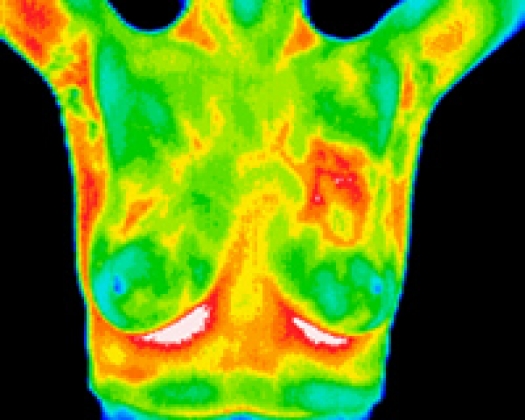
Finally, consider having a Breast Thermogram.  This is a form of Breast assessment that does not require exposure to radiation.  Instead, it measures the heat released from the Breast, which indicates areas of increased cellular activity/growth.  A Thermogram can identify problems in Breast Tissue years before they can be seen by a mammogram or felt by your doctor.  

Other Important Steps to Take Include:

  • Reduce the use of pesticides, pollutants, and chemicals in your home
  • Eat organic foods that are free of hormones and pesticides
  • Maintain an ideal body weight (excess fat increases estrogen production)
  • Exercise regularly
  • Find ways to better manage your stress


Are you at high risk for Breast Cancer?

Are you at high risk for Breast Cancer?

Signs you may be at high risk for Breast Cancer

In order to determine what the most appropriate intervention strategy for you to employ, we often recommend testing. Early detection and correction of risk factors can be one of your biggest allies in reducing your risk. If any of the following apply to you, you may be at high risk:

Excess Iron

How to test: Have the recommended Blood Tests coupled with a HTMA

Common in women after they stop menstruating, elevated iron levels can be indicative of a heightened cancer risk. Excess iron in the body works as a powerful oxidant, increasing free radicals thereby raising your risk. If you are a post-menopausal woman or have been diagnosed with breast cancer you will certainly benefit from having the recommended tests above. With these test we can establish if your iron recycle system is working properly. If the iron recycle system is not working, increased oxidative stress will occur in the body, increasing your risk of Breast Cancer, inflammation and indeed any other disease.

Estrogen Imbalance

Researchers have found that the body metabolizes estrogen into several different metabolites that can impact cancer development. One metabolite, 2-hydroxy-estrone, tends to inhibit cancer, whereas another 16-a-hydroxy-estrone can actually stimulate tumor development.A woman’s “bio-mechanical individuality” and liver metabolism determines which of these metabolites predominates. As iron accumulates in the body, this is a sign of low FOX enzyme function , (this is the master antioxidant enzyme). and estrogen is increasing as a direct result. The liver detoxifies estrogen in two different phase 1 phase 1 & phase 2. The results of the Tests above will provide information regarding the liver function. We will be specifically looking at the minerals that run the liver, deficiencies or mineral imbalances will be addressed..

Elevated Breast Inflammation

How to test: Get a Breast Thermography
A Breast Thermography is an assessment tool that looks at the function of the breast tissue. Remember change in function happens 8 to 10 years before a cancer will be detected on a structural test such as a mammogram or ultra sound.. By identifying areas of high inflammation, you can address the underlying cause of the inflammation before cancer has a chance to develop.

What else can you do to REDUCE your risk?

The steps we recommend taking to reduce your risk of Breast Cancer are also those of reducing your risk for many other diseases. In an effort to help you live a healthier life, we invite you to take note of which areas of your life need a little extra attention. Even small changes can lead to healthy habits, and can dramatically impact your future health.

Eat a wholefood diet

It is important to address your diet, This will nourish your body, aid in detoxification, and support the following breast cancer prevention strategies. Specific mineral and enzyme deficiencies will be identified through the above tests and addressed to restore balance and healing to your body.

Maintain a healthy balance of Estrogen

Estrogen is produced in fat tissue, which is the primary non-ovarian site for production. Consequently, women with excess weight have higher levels of estrogen which are associated with a greater risk for breast cancer. It is imperative that you maintain a healthy body weight if your goal is reducing your risk.We recommend and believe in reducing your weight and balancing your hormones, by adopting a nutritious diet and engaging in regular, physical activity to help prevent breast cancer.

Apply this advice today by structuring your week, so you log 30 minutes of exercise on most days of the week. If you have a hard time getting a solid 30 minutes, try breaking up your workouts to two shorter 15 minute bouts. When it comes to weight loss and exercise, weight training can have the greatest impact on your metabolic rate. In other words, when you build muscle, you’ll burn more fat calories throughout the day (even when you aren’t exercising). Over time, building muscle can have a big impact not only on losing weight but maintaining it.

Reduce Stress

Many studies have shown long term stress has a very negative impact on the body. Chronic stress has a profound impact on the liver biochemistry and enzyme function. It is important as part of your Healing Journey you identify, and find ways to reduce your stress. Consider such thigs as yoga, gentle exercise, meditation, EFT etc….

Get Proper Sleep

Proper sleep relies on getting enough sleep and sleeping between certain hours. According to Ayurvedic medicine, the ideal hours for sleep are between 10 pm and 6 am. Modern research has confirmed the value of this recommendation, as certain hormone fluctuations occur throughout the day and night. If you engage in the appropriate activities during those times, you’re ‘riding the wave’ so to speak, and are able to get the optimal levels. Working against your biology by staying awake when you should be ideally sleeping or vice versa, interferes with these hormonal fluctuations. You are at an increased risk of developing Breast Cancer ?

Our approach when treating a person is to establish the Root Cause of illness or disease. The body is a Self Healing Machine We need to identify and remove the obstacles in it’s way, by balancing, nourishing and supporting the body.

What the Doctors are saying about Thermography.


“Every woman should include Breast Thermography as part of their regular breast heath care. I have recommended the use of this technology extensively over the years in my newsletter. Thermography has the unique ability to “map” the individual thermographic readings in the breast.. Christiane Northrup, M.D. Noted Author and Leading Expert in Women’s Health Two years ago I researched Breast Thermography for an article in my newsletter and I was impressed with what I found. Finally, there was a sensitive test to support the information from mammograms. In addition, it could also detect potential problems at a very early stage. For years, I’ve been looking for a test to offer my patients who refuse mammograms. Some are concerned about cumulative radiation and complications from compression, and others have personal reasons for refusing this test. Yet, to do nothing is to put their heads in the sand, and this doesn’t save lives.
With all this information in hand, I decided to find out first hand what this technology was all about. I contacted Dr. Amalu and he spent some time going over the research and background of Breast Thermography. I also decided to have a Breast Thermogram. I was pleasantly surprised with the comfort and ease of the examination. I found in my research, and personally, that Thermography can help a woman take a possible
precancerous condition and turn it around. It supports mammography and provides additional information. Prevention is the gold standard of health care. In my opinion, Breast Thermography should be part of every woman’s yearly exam to help prevent breast cancer, and for early detection.
I will continue to have regular Thermograms and intend to keep writing about it in my newsletter until there are centers throughout the country.


Scott T. Miles, M.D., F.A.C.O.G., C.T.T. Fellow American College of Obstetrics and Gynecology. Women need to know that Breast Thermography is a promising and safe technology that is a welcome addition to helping women create breast health.


Susan M. Lark, M.D. Distinguished Author and Leading Expert in Women’s Health:
Breast Thermography is an excellent adjunctive arrow in the quiver of tools for breast screening. Whereas structural screening looks for static anatomical abnormalities, Thermography reveals physiological changes in the breast allowing for earlier intervention in the detection of remalignant
and malignant processes. Additionally, Breast Thermography allows for proactive rather than just reactive measures in the fight against breast cancer. However, it is not intended to replace structural screening, but rather is an extremely valuable addition to a Mammogram.


Nan Kathryn Fuchs, PHD Editor, Women’s Health Letter
My patients are overjoyed to find out about how Breast Thermography can help them maintain their breast health. Not only can the test pick up abnormalities long before they manifest via mammography, but it can also detect patterns of estrogen dominance, a possible precursor to breast cancer, which we can then work to change by balancing a woman’s hormone profile naturally. And it’s so simple and painless. My own experience of having a Thermogram was very comfortable and took about 30 minutes. Multiple images from different angles were taken of my
breasts and digitally processed by a sophisticated computerized system. I had had a recent breast ultrasound, wherein they could barely find the small superficial lump I had discovered. It was reassuring to add Thermography to my other tests for a more comprehensive examination.
I feel that is it important for every woman to have regular breast Thermograms. Every new patient is informed about Breast Thermography and I provide them with a brochure that explains the procedure.


Carol Knight, M.D. Private Practice Women’s Health:

The use of computerized medical infrared imaging for breast cancer detection, diagnosis, and as a high risk and prognostic indicator leads to both earlier detection of breast cancer and increases the overall survival of breast cancer patients.


Robert Elliot, M.D., Ph.D. Comprehensive Breast Care Specialist, Founder and Director EEH Breast Cancer Research and Treatment Center, President American Mastology Associationn. far Infrared imaging of the breast should be an integral part of any breast cancer screening program due to its value as an independent risk factor for breast cancer and its value as a prognostic indicator.


Jonathan Head, Ph.D., Tumor Cell Biologist and Pioneer in Breast Cancer Vaccines Director of Research EEH Breast Cancer Research and Treatment Center, Associate Professor of Biochemistry Tulane University:

Thermography imaging, based more on process than structural changes, and requiring neither contact, compression, radiation nor venous access, provides pertinent and practical complimentary information to both clinical examination and mammography. Quality controlled abnormal infrared images heighten our index of suspicion in cases where clinical or mammographic findings are equivocal or nonspecific and signal the need for further investigation rather than observation. With the addition of infrared imaging, our sensitivity of image detection has increased from 83% to 93%.


John Keyserlingk, M.D., Ph.D. Oncological Surgeon, Ville Marie Breast and Oncology Center, Department of Oncology St. Mary’s Hospital, Montreal, Quebec

Abnormal Breast Thermogram is the highest known risk marker for the presence of or future development of breast cancer. As such, Breast thermography should be a part of every woman’s regular
breast health care.

Thermography for the Prevention of Breast Cancer

Thermography is a procedure in which a heat-sensing infrared camera is used to record the surface heat produced by different parts of the body. Abnormal tissue growth can cause temperature changes, which may show up on the thermogram. Thermography may be used to diagnose breast cancer and other tumors.

Definition from the National Cancer Institute

We are all familiar with breast cancer walks, races, fund-raisers, luncheons, etc., raising millions if not billions of dollars for research for “the cure”. Why then, in 1940 only 1 in 22 women got breast cancer, yet today it is 1 in 8? Shouldn’t we be focusing on prevention, rather than wait until we are diagnosed to do something about it? Most of us are less familiar with the tools of prevention. Mammography, MRI, ultrasounds, are all words most have heard but thermography is much less familiar.

With over 97% accuracy, thermography has the ability to warn women 8-10 years before any other imaging method, that a cancer may be forming, alerting women early enough to begin the process of actually reversing this abnormal development before it even becomes a tumor. Thermography uses NO compression and NO radiation, so it is safe, non-invasive and painless.

Thermography is designed to detect and reveal abnormal heat and vascular development in the breast tissue, which is attributed to early tumor growth. Every physician knows that a tumor, whether malignant or benign, needs its own blood supply to nourish itself to grow. These vascularities grow in very abnormal patterns, creating the heat and inflammation that a thermal imaging camera detects.

An abnormal thermogram is the single most important marker of high risk for developing future breast cancer, ten times more significant than a family history of the disease. In other words, if someone gets a suspicious thermogram indicating that something is wrong, something usually is.

Did you know that the single greatest risk factor for development of breast cancer is a woman’s lifetime breast exposure to estrogen? A woman can have up to fifty times more estrogen in her breast tissue than her blood levels indicate. With the help of a natural practitioner or qualified nutritionist, a woman can make changes to affect her hormone levels and lower her risk. Throughout this time, a woman can monitor her breast tissue changes with thermography and actually see that the changes made to correct the imbalance are working. This is where breast thermography plays an unprecedented role in breast cancer prevention.

Along with over 800 published peer-reviewed studies on the effectiveness of thermography, a study published in the January 2003 publication of the American Journal of Radiology showed thermography to be a “safe, non-invasive and valuable adjunct to (not replacement for) mammography in determining whether a lesion was benign or malignant, with over 97% sensitivity”.

Unfortunately, until thermography is considered the “standard of care,” most women won’t even hear of this life-saving technology. Every woman should have access and be aware of this valuable screening tool. It is the earliest detection available, and provides hope in the prevention of this epidemic disease.

Confirmed: Increased Bone Density, Increases Your Breast Cancer Risk

Confirmed: The Lower Your Bone Density, The Lower Your Breast Cancer Risk

When older women are told that their bones should be as dense as a young adult (30 year old) at peak bone mass, things can and do go terribly wrong.

A new study published in The Breast Journal, authored by researchers at the Department of Surgery, St. Luke’s-Roosevelt Hospital Center, New York, NY confirms numerous past studies showing low bone mineral density lowers the risk of breast cancer,[i], [ii], [iii], [iv], [v], [vi], [vii], [viii], [ix], [x], [xi],[xii], [xiii]  and validates the hypothesis that women with breast cancer and low bone mineral density will have lower breast cancer recurrence rates than women with so-called ‘normal’ bone density.

In the study, titled, “Bone mineral density is a prognostic factor for postmenopausal Caucasian women with breast cancer,” researchers reviewed the records of 309 breast cancer patients with complete follow-up and bone density tests within 1 year of their surgery. The results were reported as follows:

The outcome of patients with low bone density (t > -1.0) was compared to the outcome for patients with normal bone density (t < -1.1). Among the 193 patients with invasive breast cancers and low bone density, distant disease-free survival at 5 years was 96% compared to 84% for 114 patients with invasive breast cancers and normal bone density (p = 0.0239). Local disease-free survival was 94% for low bone density patients compared to 86% for patients with normal bonedensities (p = 0.0794).

In summation, low bone mineral density is associated with a lower local and distant rate of breast cancer recurrence.

The New Normal: Aging As A Disease

It may come as a surprise that high bone mineral density is not somehow magically healthier for overall women’s health, as the myopic focus on increasingly bone mineral density by any means necessary has become a quarter of century long medical obsession, despite the fact that it is based on a warped, if not also dangerous, prioritization of women’s health concerns.  In fact, the weight of evidence is now driving us towards the opposite conclusion: unnaturally high bone mineral density is dangerous to women’s health.

First, let us take a closer look at what so-called ‘normal’ bone density means.

Ever since the natural aging (and thinning process) of the bones was redefined as a disease by the World Health Organization in mind mid-90’s, and the young adult (T-score), based on a 30-year old woman at peak bone mass, was used to determine ‘normal’ bone mineral density, millions of asymptomatic and presumably healthy women have been overdiagnosed  and overtreated with ‘osteoporosis’ or ‘osteopenia.’

Commonsense says that you should not compare older women’s scores with that of a young adult at peak bone mass, but financial pressure to capitalize on new drug development (Merck’s bisphosphonate Fosomax) and diagnostic technologies (DXA scans) made this type of de novo disease creation a profitable business model, regardless of its veracity or ethicality.

Of course, these new definitions lead to overtreatment, both with calcium supplements and bone-targeted patent drugs, further driving bone mineral density levels higher without thought to the unintended, adverse health effects, which we now know include both heart disease and cancer. [See: Osteoporosis Myth: The Dangers of High Bone Mineral Density]

Bone Fracture Associated Death Vastly Lower Than Breast Cancer & Heart Deaths

What, in fact, the new study has found is that having age-appropriate bones (measured through the Z-score) is in fact healthier, even if the (mis)use of the T-score distorts them into being ‘low’ relative to a 30-year old woman.

Research like this points to a fundamental failing in the way conventional medicine prioritizes women’s health issues. Whereas breast cancer risk reduction is presumably of utmost importance, given the year long drive to raise ‘breast cancer awareness’ through the promotion of fundraisers and of course breast screenings – this despite the now well-known fact that over 70,000 women a year are misdiagnosed and wrongfully treated for ‘breast cancers’ that weren’t actually anything beyond natural variations in breast morphology that would never progress to cause harm, much less symptoms in the majority of women – the fact remains that women are being corralled into receiving bone mineral density scans that reinforce the false perception that a woman will be overall healthier, and mortality lower, if only her bones were denser than is normal for her age.  Nothing could be further from the truth…

First, women die today primarily from heart-related deaths, such as heart attack associated with coronary artery disease. The drive to increase bone mineral density results in women taking artery-calcifying and heart-stopping megadoses of 1000 mg or more daily of inorganic calcium, even when as little as 500 mg a day of elemental calcium can increase heart attack risk by 24-86%, according to a series of meta-analyses published last year.

Second, after heart disease, cancer is the second highest cause of death in women, with breast cancer mortality second only to lung cancer mortality.

Juxtaposed to heart- and cancer-related causes of mortality in women, accounting for 24% and 22%, respectively, lethal complications from fractures associated with low bone mineral density do not even figure into the top thirty causes.[xiv]

So, the shattered, fractionated medical system grinds on. The ‘bone doctor’ creates the impression in women that nothing could be more important to women’s health than increasing bone density, even if this obsessive drive results in increased breast cancer risk and poorer prognosis, and calcified arteries from megadose calcium supplements.  The ‘breast doctor’ focuses on diagnosing breast cancer early, planting seeds of radiation-induced cancer into their bodies, while misdiagnosing hundreds of thousands of women. The ‘heart doctor’ suppresses LDL cholesterol via chemical poisoning (statin drugs), resulting in the weakening of their patients’ heart muscles.

Sheer madness masquerading as the standard of care.

error: Content is protected !!