News

Breast Thermography: What you need to know


Breast Thermography is a non-invasive and painless test, with no radiation involved. It can detect and monitor early warning signs of breast dysfunction and inflammation.

This type of breast cancer screening is particularly useful for people under the age of 50. This is because mammography, another type of screening, can be less effective for this group.

Thermography is approved by the FDA since 1982 for use a s an adjunct to to mammography. and .

How Thermography works

Thermography, also known as thermal imaging, detects the rise in skin temperature which may occur when cancer cells are multiplying.

The way Thermography, or thermal imaging, works is based on how cancer cells grow. When cancer cells are growing and multiplying in a tumor, blood flow is very fast in that area.

Increased blood flow makes skin temperature increase. This rise in skin temperature is what breast thermography is aiming to detect.

There is a debate among doctors about how useful Thermography is as a way to diagnose breast cancer. The American Cancer Society have said that it will take time to see if it is better than, or equal to, current tests.

Process of a Thermograph

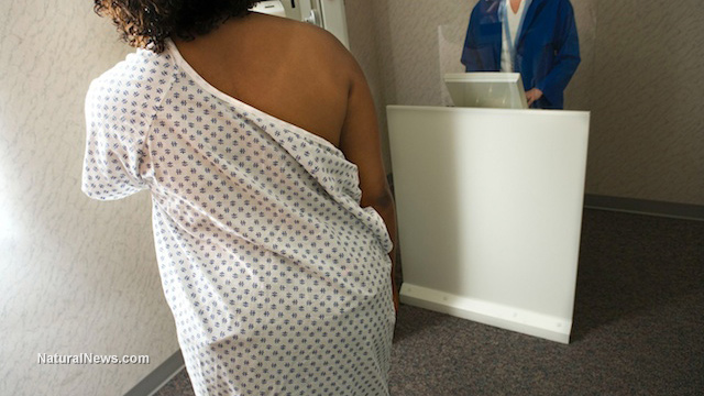
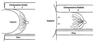
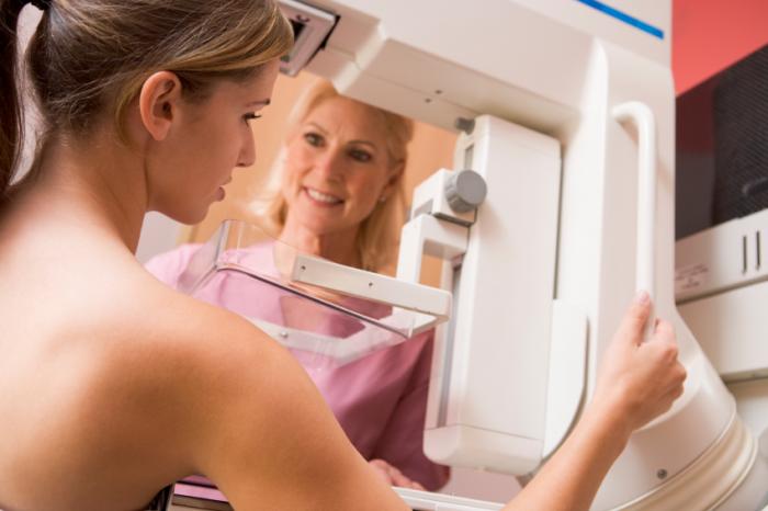
Breast Thermography is a non-invasive physical test.. It is also “non-compressive,” which means that it does not put force on, or squeeze, the breast, as is the case with breast mammography exams.

Some people are worried about the force put on breasts in a mammogram, so prefer the idea of a Thermogram.

Thermography uses digital infrared imaging to detect subtle changes in the breast based on symmetry. It looks for clear abnormalities in one breast in comparison to the other. This has to be adjusted when using on individuals who have undergone a mastectomy

To understand a Thermography report, it is necessary to know two things about cancerous breast tissue, compared to normal breast tissue. These are:

  • there is more metabolic activity (biochemical reactions)
  • there is increased blood flow

These aspects of breast cancer tissue result from the cancer cells doing all they can to maintain and grow. Another side effect of this is a rise in skin temperature.

Ultra sensitive cameras and computers can detect this increase in temperature. They produce high-resolution images.

Using thermography with other tests

woman having mammogram

T

Thermography can be used alongside other tests, such as mammographic or ultrasound screening.

Normally if a Thermograph alone is used, the images taken will be kept on record and used for future evaluations. The idea is that an initial Breast Thermography Test, which can be used on people as young as 18, will provide a baseline.

Future tests can then be compared to this baseline to see if there are any changes or abnormalities that develop.

Follow up tests

If abnormalities are detected, then follow-up procedures will be required to investigate further. This may include a mammogram.

These follow-ups can also rule cancer out, as the images could be showing a host of other breast diseases. When abnormalities are present it could be a sign of:

  • cancer
  • fibrocystic disease
  • infection
  • vascular disease

Results categories

After the test, the reports are divided into five categories. These is known as the TH (thermobiological) grading system. The categories are as follows:

  • TH-1: Symmetrical, bilateral, nonvascular (non-suspicious, normal study)
  • TH-2: Symmetrical, bilateral, vascular (non-suspicious, normal study)
  • TH-3: Equivocal (low index of suspicion)
  • TH-4: Abnormal (moderate index of suspicion)
  • TH-5: Highly abnormal (high index of suspicion)

Follow-up exams are needed at different times for each category, a rough guide is as follows:

  • TH-1 and TH-2: every year
  • TH-2: every year
  • TH-3: every 6 months
  • TH-4 and TH-5: every 3 months

Effectiveness

People do not have to decide between breast thermography or mammography, but may use them both. Using the two together can be more effective.

The use of Thermography, Mammography, and a clinical exam is known as the “multimodal approach”. Using this approach can help identify around 95 percent of early stage cancers.

Technology

Thermography camera

Thermography uses sophisticated cameras and computers to capture infrared photographs.

Thermal imaging technicians capture an infrared photograph, or heat image, of the breast.

This can then be printed in high resolution for a doctor to study, or may be sent to them electronically.

History

Thermography has been experimented with in medical science for many hundreds of years. However, it wasn’t until 1972 that the Department of Health, Education and Welfare announced that Thermography was “beyond experimental.”

This announcement applied to the use of the technology for many areas, including the evaluation of the breast. The advance in technology since then has seen Thermography become recognized in multiple areas of medicine, including breast health.

Heat patterns

The cameras used in Breast Thermography produce ultra-sensitive, high-resolution infrared images. These images show heat patterns and identify changes in the temperature of the skin and blood flow.

Many other technologies that test for breast cancer work in other ways that do not involve looking for heat patterns. These tests include:

Breast thermal risk index

Other factors can affect the results of a Breast Thermography. So, often doctors will use the Breast Thermal Risk Index to help ensure more accurate results. This includes:

  • age of person
  • family history of breast cancer
  • medication, including birth control and hormone pills
  • menopause
  • if the person is overweight

Benefits

Thermography offers the opportunity for early detection of breast cancer and has a number of specific benefits.

Earliest possible detection

A Thermogram enables abnormalities to be detected sooner than other procedures.

Compared with other procedures being used on their own, Thermography makes it possible for cancer or pre-cancerous growth to be detected up to 10 years sooner than they may otherwise have been.

Close monitoring

Thermography means that people at potential risk of developing breast cancer can be monitored closely.

Outlook

If breast cancer is caught at an early stage through the use of thermographs, this increases a person’s treatment options and should ultimately lead to a more positive outlook.

However, Breast Thermography only has the potential to identify early warning signs. Thermography, mammography and ultra sound cannot diagnose breast cancer, Only a biopsy can do that, Thermography is the only assessment tool that may highlight a potential problem years before a lump is detected on other structural tests. Thermography is truly about Breast cancer Prevention, a health and wellness Programme can be followed to restore Breast Health. Again the use of Thermography will identify the effectiveness of such a programme.

<

Surgeons admit that mammography is outdated and harmful to women


Breast cancer

(NaturalNews) Every year, millions of women flock to their doctors to get their annual mammograms, a breast cancer screening procedure that involves pressing a woman’s breasts between two metal platforms to scope out tumors. But surgeons everywhere are starting to question the controversial practice, which studies show isn’t even an effective screening tool, and is actually harmful to the bodies of women who receive it.

The public is told that mammograms are the only way to catch breast cancer early, but a review of eight scientific trials evaluating the procedure, found that mammography is neither effective nor safe. After looking at data on more than 600,000 women between the ages of 39 and 74 who underwent the procedure on a routine basis, researchers found that many women are misdiagnosed. Many of these same women are consequently mistreated with chemotherapy, resulting in their rapid demise.

As published in the Cochrane Database of Systematic Reviews, the review concluded that mammography causes more harm than good, because many more women end up being misdiagnosed and mistreated than those actually avoiding the development of terminal breast cancer. Thus, the procedure known as mammography is an outdated scourge that belongs in the history books of failed medical treatments, and not at the forefront of women’s medicine.

“If we assume that screening reduces breast cancer mortality by 15% and that overdiagnosis and overtreatment is at 30%, it means that for every 2000 women invited for screening throughout 10 years, one will avoid dying of breast cancer and 10 healthy women, who would not have been diagnosed if there had not been screening, will be treated unnecessarily,” the authors concluded.

Group of top medical experts admits mammography does more harm than good

One year after this review was published, a second one published in The New England Journal of Medicine (NEJM) came to a similar conclusion. A team of medical professionals that included a medical ethicist, a clinical epidemiologist, a pharmacologist, an oncologic surgeon, a nurse scientist, a lawyer and a health economist, decided that the medical industry’s claims about the benefits of mammography are essentially bunk.

They found that for every 1,000 women screened in the U.S. over a 10-year annual screening period beginning at age 50, one breast cancer death would be prevented, while a shocking 490 to 670 women would have a false positive, while 70 to 100 would undergo an unnecessary biopsy. Between three and 14 of these women, the study found, would also be over-diagnosed for a non-malignant form of cancer that never even would have become “clinically apparent.”

This study out of Switzerland corroborates another out of Canada – the 2014 Canadian National Breast Screening Study – which concluded in lockstep with the others that mammography screenings do not reduce mortality rates from breast cancer any better than a simple physical examination. In other words, the procedure is completely unnecessary, and in many cases exceptionally harmful.

And on and on the list goes, with data out of Norway and elsewhere confirming that mammography isn’t all that it’s cracked up to be. U.S. data spanning the course of nearly 40 years shows that more women are over- or misdiagnosed with breast cancer because of mammograms than are successfully early-diagnosed with breast cancer in such a way as to protect against metastasization. This represents an exceptionally poor track record that calls into question why mammography continues to be used when it clearly doesn’t work.

“I believe that if you did have a tumor, the last thing you would want to do is crush that tumor between two plates, because that would spread it,” says general practitioner Dr. Sarah Mybill, as quoted in the documentary film The Promise.

Sources for this article include:

NaturalBlaze.com

NCBI.NLM.NIH.gov

NEJM.org

THERMOGRAPHY OVER MAMMOGRAPHY FOR BREAST CANCER SCREENING

thermography for breast cancer screening

THERMOGRAPHY OVER MAMMOGRAPHY FOR BREAST CANCER SCREENING

If cancer is caught in it’s early stage, there is a better chance of treating it successfully. And, because at at early stage, it has less likely to spread, a simple surgery may be able to remove them.

Latest research works have been successful in favoring thermography for breast cancer screening for early detection. Here is how it is emerging as a promising technique for the upcoming years:

Tracing heat patterns of the breast cells can predict the course of the disease as well as the risks quite earlier than its outcome. A study was conducted that involved 1,245 women who underwent:

  • physical examination
  • ultrasonic investigation
  • mammography
  • fine needle aspiration
  • biopsy for breasts

One-third of the subjects showed up with histologically-proven cancers within a time frame of five years. In their cases, thermography was able to detect metabolic changes in the lesion cells that had the potential to grow rapidly, and its predictive value remains far better than that of a biopsy.

When opting for a procedure, you would certainly want to decide in favor of the one that yields accuracy of high scores. Experimental studies have been successful in concluding that cell patterns exhibited on a thermogram are nearly 80 percent accurate. Hence, thermography takes an edge over mammography.

With mammogram, it may be harder to peak through the anatomical abnormalities of denser breast tissues, especially those of premenopausal women.

On the other hand, in a study that compared efficacies of the three most-adopted breast cancer screening modalities: clinical examination, mammography, and thermography; the certainty of the thermography for larger lesions and for the ones with lymph node metastasis was significantly lower as compared to that of mammography.

A compelling reason that questions the role of mammography is the high probability of “false positive results”. A retrospective cohort evaluation for breast cancer screening and diagnosis of 2,400 women subjects falling between the ages of 40 and 69 years led to the discovery that, mammography stood for a cumulative risk of 49.1 percent of false positive results on an estimate. The research included four mammograms and five clinical breast examinations per subject over a 10-year period.

5 ADVANTAGES OF OPTING FOR BREAST THERMOGRAPHY SCREENING

1. Suitable for younger women

It should be the first choice for premenopausal women whose breasts tend to be denser. Thermography can differentiate fibrocystic tissue, scars or breast implants from that of cancerous cells.

2. Detect cell changes outside of the breast region

Many times precancerous cells can start forming from the armpit area due to use of underarm deodorants, and the concentration of lymph nodes in that area. Mammography is not the best method to use for screening outside of the breast region, the armpit area included.

3. Great as an additional test

Whatever method you decide to use, thermography will be a great additional test due to its accuracy and early detection ability. Should you discover a lump from your self-examination, a thermography will be able to give you a more conclusive diagnosis as to the type of lump/growth it is.

4. It is painless and radiation-free

As you may have experienced or heard from other women, the pressure of a mammogram machine is equivalent to putting a 50-pound weight to compress your breasts. Many who have done mammogram will be able to tell you that it is painful. And this is especially traumatic for women who have small breasts.

It doesn’t make sense to use high exposure to radiation from mammography to find what we know is caused by radiation!

With thermography, you will be required to sit in front of the scanner that scans your body heat. Thermography is non-contact, non-invasive, uses no painful breast compression, nor emits any harmful radiation.

5. It is safe

Thermography is so safe that it can be used on pregnant and nursing women without any side effects whatsoever. It’s merely taking an image of your body heat, like shooting a photo!

IS THERMOGRAPHY AVAILABLE IN YOUR COUNTRY?

Thermography Clinic Ireland is located in : Suite 1, Portmarnock Business Centre, Strand Road, Portmarnock, Co Dublin, Ireland

CONCLUSION ABOUT THERMOGRAPHY

As with anything so important as your health, I suggest that you make an informed decision. Know the source of your information. There will always be two sides to healthcare. There is the conventional, invasive method; and there is the traditional, holistic and natural approach. Decide which route you wish to take.

You may have been told or formerly educated about mammogram but now decided that it is, after all, not the best way to go. There is no need to feel any guilt nor be intimidated into going that route. Take your own health into your own hands and decide what you think is best and safe.

When done properly, thermography for breast cancer screening has the true potential to detect breast cell anomalies long before mammography can detect cancer. By early detection (up to eight years earlier), it allows you to get help and implement lifestyle changes that can improve your health significantly.

Breast Implants – Mammography and Thermography

Image result for breast implants

Approximately 4 million women have had breast implant surgery in the U.S.  Many do not realize that typically silicone or saline implants have a reliable life of 10 years.  The FDA warns that after 10 years at least one implant is likely to rupture and by twenty years both implants will have ruptured.  This indicates that implants should be monitored for leaks and ruptures and replaced or removed periodically to protect her health.  If the saline or silicone is not sterile and leaks into the body, it can an often does, cause irreparable damage to her health.  Fibromyalgia can be directly related to such events.

The FDA reports that mammography screening will most likely reduce the life of the implant, and ruptures during the procedure do occur.  Even so, annual mammogram screening is recommended for women with implants, although sensitivity is affected.  The false negative rate increases from 33% for women without implants to 55% for women with implants.  For these reasons, women with implants may choose to avoid mammography.

This image has an empty alt attribute; its file name is Breast-Implant.jpg

Paragraph

 For those women who still desire to access their breast health, Thermography is a safe and worry-free method, as there is no possibility of directly affecting the implants.  Also, women are expected to have a baseline screening before implant surgery.   Many are too young to be exposed to the radiation of x-ray and screening with Thermography would be a wise choice.

Is There a Link Between Estrogen Dominance and Breast Cancer?

Posted on November 17, 2015

Is There a Link Between Hormonal Imbalance and Breast Cancer?

There is a clear increase in a woman’s chances of getting breast cancer as they age, in particular once they get into their 40′s, 50′s and beyond. Why would there be such an increase in breast cancer rates in women at this age? What is the significance of this time in a woman’s life?

Hormone Imbalances and Estrogen Dominance

During this time, a woman is likely experiencing hormonal imbalance as she enters peri-menopause, and eventually menopause. The primary reasons for this imbalance are:

  • declining levels of progesterone that occurs with aging, surgical removal of the ovaries or excessive stress (high cortisol equals low progesterone)
  • higher levels of estrogen as the body struggles make an egg from aging ovaries
  • accumulation of environmental hormone disruptors in the body from a lifetime of exposure to pollutants, plastics and pesticides (all contain xeno-estrogens)
  • use of oral contraceptives (past or current)
  • synthetic hormone replacement therapy
  • an unhealthy digestive tract (where hormones are removed from the body)

These factors can contribute to an excess of estrogen in the body known as Estrogen Dominance.  Since estrogen stimulates cell growth, a predominance of it, especially in the absence of adequate levels of progesterone, presents an increased risk of cancer, particularly in the breast.

Don’t get me wrong, estrogen is not a demon!  It is all about balance.  To better understand, let’s explore the relationship of estrogen to cell growth, particularly in the breast.  Humans produce three types of estrogen: Estrone, Estradiol and Estriol.  Each has a different effect on breast tissue:

  • Estradiol: The most potent estrogen, equally stimulates and inhibits cell growth. In other words it is neutral.
  • Estrone: The second most potent estrogen, stimulates cell growth.
  • Estriol: The weakest estrogen, inhibits cell growth.

The key takeaway here is that having levels of Estrone that are too high in your body may result in more breast stimulation.  While higher levels of Estriol in your body results in less breast cell stimulation, and therefore, breast protection.

But I’ve Heard That Hormone Replacement Therapy (HRT) Increases Your Risk of Breast Cancer?

You may have heard that hormone replacement therapy causes breast cancer.  Well, if the patient is taking Premarin or other hormones, then that would actually be true. The ugly fact is that Premarin and other such synthetic hormones are composed of 48% Estrone (the potent stimulatory Estrogen). In a nutshell: you are taking more of the bad estrogen and decreasing your amount of the good estrogen.  This equates to a prime set up for breast cancer.

What About Progesterone?

Progesterone is a key component in helping to prevent breast cancer.  Progesterone is the dominant hormone in the second half of a woman’s cycle.  It arrests cell growth and causes the shedding of the uterine lining.  In the breast, it prevents fibrocystic changes and encourages apotosis (programmed cell death).  Without enough progesterone, women may experience heavy periods, uterine fibroids, endometriosis, breast cysts and cancer!

Basically Estrogen = GROWTH and Progesterone = NO GROWTH.  Without adequate amounts of Progesterone to balance the stimulatory effects of the estrogen go unchecked. This over stimulation is particularly bad for the breast tissue.

Progesterone vs. Progestin:

Progestins are a classification of drug that are similar to the natural progesterone you produce in your body.  However, just as the name is similar but not identical, neither are their molecular structures.  The Women’s Health Initiative revealed a 26% increase in breast cancer as a result of taking synthetic progestin.  The Nurse’s Health Study found that synthetic progestins tripled breast cancer risk over that of estrogen only.  The use of Provera, a synthetic progestin and component of Prempro, has been shown to increase the risk of breast cancer by 800%.

Progesterone has many Anti-Cancer Benefits:

  • Progesterone decreases estrogen production
  • Progesterone moves estrone to an inactive (sulfated) form
  • Progesterone moves estrone to weakest/safest estriol
  • Progesterone down-regulates estrogen receptors all together
  • Progesterone activates the cancer protection gene, p53

Simply stated, progesterone is breast protective.

Know The Symptoms of Hormone Imbalance:

  • Unwanted weight gain/inability to lose weight
  • Premenstrual weight gain/bloating
  • Heavy/painful periods
  • Irregular bleeding
  • PMS
  • Tender and/or fibrocystic breasts
  • Mood swings
  • Low Libido
  • Depression
  • Low thyroid symptoms
  • Sleep disturbances
  • Fatigue
  • High stress
  • Belly fat
  • Low immunities/susceptibility to illness

What Can I Do to Prevent / Treat Estrogen Dominance?

First, I recommend assessing the root cause of hormonal imbalance. l use HTMA and an a specialised Blood Test to determine the extent and the root cause of the estrogen dominance. This will give them the best picture of your unique situation and needs.  

Estrogen Dominance is a pandemic on the planet it is as a result of mineral dysregulation. Women with low bioavailable copper cannot make enough of the body’s master antioxidant feroxidase so the body goes to plan B and increases the production of estrogen to use as an antioxidant to offset the oxidative stress in the body. This is the very beginning of hormonal imbalance.

Consider having a thermogram.  This is a form of breast asessment that does not require exposure to radiation.  Instead, it measures the heat released from the breast, which indicates areas of increased cellular activity/growth.  A thermogram can identify problems in breast tissue years before they can be seen by a mammogram or felt by your doctor.  We are located in Portmarnock, Co Dublin.  To book an appointment, simply call our office at Ph: 086 1623683.

  • Reduce the use of pesticides, pollutants, and chemicals in your home
  • Eat organic foods that are free of hormones and pesticides
  • Maintain an ideal body weight (excess fat increases estrogen production)
  • Exercise regularly
  • Find ways to better manage your stress
  • For more information about managing estrogen dominance, hormone assessment, including adrenal and thyroid assessment. Contact Thermography Clinic Ireland.  Ph: 086 1623683 or e mail info@thermographyireland.ie

Take a Step Towards Better Health

A

error: Content is protected !!